


Overview of Lung Fibrosis and Importance of Early Detection
Lung Fibrosis encompasses various chronic lung diseases, primarily resulting in lung tissue damage and scarring. This condition leads to a progressive decline in lung function, with symptoms including shortness of breath and persistent cough. Common types include idiopathic pulmonary fibrosis and connective tissue disease-associated interstitial lung disease.
Statistics highlight the prevalence of lung fibrosis as increasing, with idiopathic pulmonary fibrosis (IPF) affecting approximately 3 million individuals worldwide. The outcomes associated with advanced stages are often grim, leading to a significant reduction in life expectancy and quality of life.
In parallel : Personalizing Treatment for Shift Work Sleep Disorder: Insights from UK Sleep Specialists
Early Detection is crucial in altering patient prognoses positively. Identifying the disease at an initial stage allows for timely intervention, slowing the disease’s progression and improving survival rates. Methods such as imaging and laboratory tests are pivotal in the early identification of lung fibrosis. Quick diagnosis can provide patients the opportunity for more personalised treatment plans, potentially involving medication, lifestyle adjustments, and, in certain cases, lung transplantation.
The vital aspect of early detection lies in the improved management of lung fibrosis, aiding healthcare providers in delivering better-targeted therapies that may enhance patients’ overall wellbeing and extend their lifespans.
Topic to read : Navigating Exotic Pet Health: Strategies for UK Veterinarians in Diagnosing and Treating Imported Animal Diseases
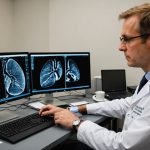
Cutting-Edge Imaging Techniques Utilized by UK Pulmonologists
The role of imaging techniques in lung health is paramount, particularly in diagnosing and understanding lung fibrosis. With advancements in diagnostic imaging, pulmonologists in the UK have embraced cutting-edge technology to enhance the detection and evaluation processes.
High-Resolution Computed Tomography (HRCT)
High-Resolution Computed Tomography (HRCT) stands out as a pivotal tool in detecting lung fibrosis due to its exceptional ability to provide detailed images of lung tissues. HRCT offers incomparable precision, allowing for the early identification of fibrotic changes even before the onset of noticeable symptoms. According to current guidelines, HRCT is recommended as a first-line imaging method in suspected cases of lung fibrosis, offering clinicians a clear route for diagnosis and helping to tailor intervention strategies promptly. Its non-invasive nature and detailed imaging capabilities make HRCT an indispensable part of modern diagnostics.
Magnetic Resonance Imaging (MRI) Developments
Recent developments in Magnetic Resonance Imaging (MRI) technology have demonstrated significant promise in lung assessment. With no exposure to ionising radiation, MRI offers a safer alternative while still providing detailed imaging necessary for detecting early signs of fibrosis. Though still emerging in the realm of lung fibrosis diagnosis, the evolution of MRI techniques suggests a future where these scans may become routine in establishing precise and safe lung health assessments.
Ultrasound Innovations
Innovations in ultrasound technology are transforming the landscape of lung diagnostics. Ultrasound imaging provides a non-invasive and radiation-free option, making it particularly appealing for assessing lung conditions such as pleural effusions and pneumothorax. While not traditionally used for detecting lung fibrosis, its utility in evaluating related symptoms and complications cannot be overlooked.
Limitations of ultrasound include its dependence on operator skill and difficulty in penetrating air-filled structures like lung parenchyma. However, ongoing advancements aim to enhance its accuracy and applicability in lung health, potentially broadening its role in fibrosis diagnosis. These innovations may soon push boundaries, offering real-time monitoring capabilities that align closely with patient needs.
Future potential sees ultrasound technology positioned to complement more established imaging techniques. Developments in portable ultrasound devices could revolutionise remote diagnostic capabilities, especially in rural or underserved regions. As technology progresses, we anticipate it becoming a staple in comprehensive lung health assessments, providing a quick, safe, and cost-effective option for clinicians in various settings. Thus, ultrasound’s role in lung diagnostics, though still evolving, promises greater support in delivering holistic care for individuals with lung fibrosis.
Laboratory Tests and Biomarkers in Lung Fibrosis Detection
In the realm of lung diagnostics, laboratory tests and biomarkers play a crucial role in improving the early detection of lung fibrosis. Blood tests are often a frontline tool, providing insight through key biomarkers like surfactant proteins and Krebs von den Lungen-6 (KL-6), which correlate with lung damage and fibrosis. As knowledge advances, these tests enhance their ability to offer early detection potential, supporting targeted interventions.
Blood Tests and Their Diagnostic Value
Blood tests carry immense diagnostic value, utilising biomarkers that indicate lung injury, aiding in predicting disease progression. The ability to identify elevated levels of specific proteins linked to fibrotic processes provides a non-invasive avenue to suspect lung fibrosis early.
Bronchoalveolar Lavage (BAL) Analysis
BAL analysis involves sampling lung secretions via bronchoscopy, highlighting its role in lung diagnostics by identifying inflammatory cells and mediators associated with fibrosis. This procedure offers detailed insights into lung health, reinforcing diagnostic accuracy.
Genetic and Molecular Testing
Advancements in genetic and molecular testing reveal susceptibility to lung fibrosis, allowing biomarkers to guide personalised approaches. Research continues to explore molecular markers, propelling the future directions of transparent and precise diagnostics for timely intervention.
Integration of Advanced Diagnostic Techniques
In the field of lung fibrosis, an integrated approach combining various diagnostic techniques is gaining significance. Diagnostic integration is essential to offer a more holistic perspective on the patient’s condition. By combining imaging methods like HRCT and MRI with laboratory tests such as blood tests and BAL analysis, healthcare providers can craft more detailed diagnostic profiles. This synergy not only bolsters patient care but also enhances the accuracy of diagnoses.
A holistic approach enables clinicians to pinpoint specific characteristics of lung fibrosis, facilitating targeted interventions. Imaging delivers precise visuals, while laboratory tests reveal molecular insights. Together, they provide a comprehensive picture, fostering timely and effective treatment strategies.
Case studies demonstrate the potency of integrated diagnostics. For instance, instances where early detection was achieved through combined imaging and biomarker analysis highlight significant improvements in patient outcomes. These cases illustrate that early intervention, guided by a robust integration of diagnostic techniques, can lead to improved prognosis, offering patients better management of lung fibrosis.
In essence, harnessing both advanced imaging and laboratory diagnostics offers a multi-faceted view of lung health, ultimately enabling healthcare professionals to deliver more personalised and effective patient care.
Impact of Technology on Early Detection of Lung Fibrosis
The realm of lung fibrosis diagnostics has been significantly enhanced by the integration of technology advancements. Among these, artificial intelligence (AI) and telemedicine stand out for their transformative potential.
Role of Artificial Intelligence in Diagnostic Evaluation
AI in diagnostics is playing a crucial role by offering enhanced imaging analysis. Through sophisticated algorithms, AI aids in recognising patterns that are often missed by human eyes, thus increasing diagnostic precision. This technology not only accelerates the evaluation process but also improves accuracy, helping clinicians make more informed decisions. Success stories abound, showcasing how AI-assisted diagnostics have markedly improved patient outcomes by facilitating early and precise detection of lung fibrosis.
Telehealth and Remote Monitoring Solutions
Telemedicine and remote monitoring have revolutionised lung health management. These solutions enable continuous patient monitoring, providing real-time health data to practitioners regardless of location. This ability to remotely diagnose and monitor conditions like lung fibrosis ensures timely medical interventions, significantly improving patient care. However, challenges such as technology accessibility and data privacy remain, requiring ongoing efforts to address these issues.
Future Technologies on the Horizon
Emerging technologies promise to further refine lung fibrosis diagnostics. Innovations are expected to enhance existing methodologies, offering even more comprehensive and precise assessments. The future holds exciting prospects for lung health, driven by ongoing technological advancements that aim to transform early detection and patient care strategies.